
Overview
Mycoplasma species are frequent contaminants in cell culture, and can be found in both cell lines and primary cell culture. According to ATCC, between 15–35% of continuous cell cultures and a minimum of 1% of primary cell cultures are contaminated with mycoplasma. Roughly 20 distinct species have been identified in mammalian cell culture, with 8 species accounting for about 95% of contamination (Acholeplasma laidlawii, M. hyorhinis, M. salivarium, M. hominis, M. orale, M. pirum, M. arginini, and M. fermentans) (ATCC, 2026).
Mycoplasma infection of cell cultures can cause many negative effects, including altering cell signaling pathways via transcription factor activation, introducing changes that can confound your results and lead to conclusions that reflect contamination, rather than your biology (Borchsenius et al., 2020).
Importantly, mycoplasma infection can be detected in RNA-Seq data. In a 2015 survey of NCBI's RNA-Seq archive, researchers found that 11% of the studies examined showed evidence of mycoplasma contamination (Olarerin-George & Hogenesch, 2015).
To identify mycoplasma contamination in customer samples, we built a mycoplasma detection screen into our RNA-Seq pipeline. It screens for the eight species above plus an additional species that emerged from customer data (M. yeatsii). The screen flags contaminated samples and reports the result in the Sample Review table. Looking across all RNA-Seq samples from the last three months, we found that at least 4% of samples and 7% of orders from academic customers were contaminated with mycoplasma spanning six of the nine species.
Detecting mycoplasma
Mycoplasma infection is challenging to detect because it doesn't cloud culture media or shift the pH, making it effectively invisible without a dedicated assay. Mycoplasma is also resistant to most antibiotics routinely added to cell cultures. Because mycoplasma infection can persist unnoticed, cell culture repositories recommend monitoring cell cultures routinely. Three methods are frequently used: direct agar culture, indirect Hoechst DNA staining and PCR-based testing.
In practice, routine monitoring is often not enough, and a culture can become contaminated between scheduled checks. Sequencing data offers an additional layer of protection: every data set is also an opportunity to screen for contamination.
Our mycoplasma detection screen
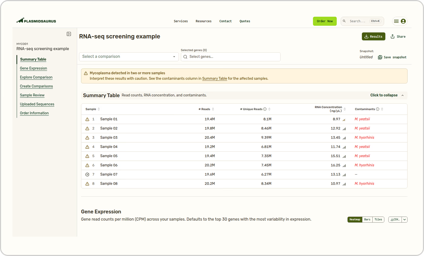
Given the impact of Mycoplasma on interpreting the results from RNA-Seq experiments, we have developed a screen to directly detect mycoplasma contamination in customer RNA-Seq results. The screen runs as part of our standard RNA-Seq pipeline and the result is reported in the Sample Review table on the Results page for your order.
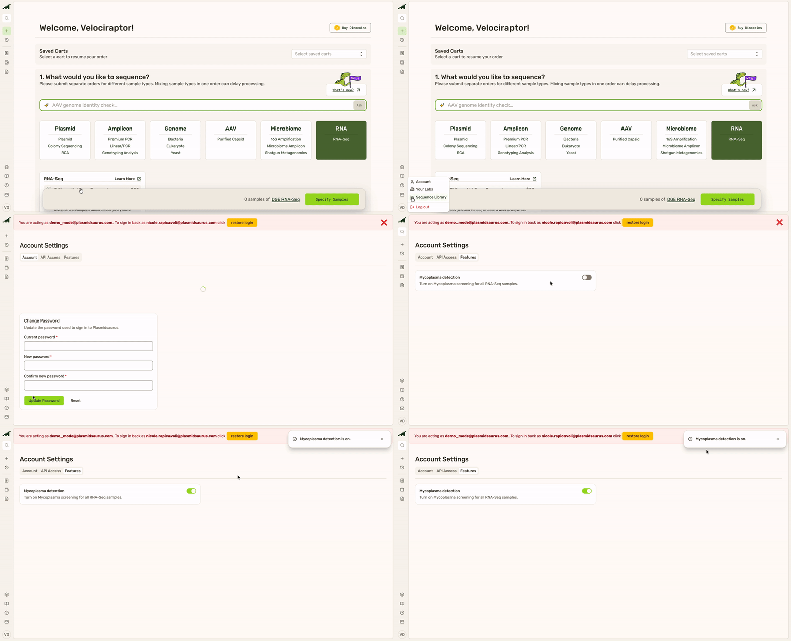
Reporting is enabled by default for academic customers. For all other customers, these results are not reported by default. This setting can be toggled on or off under Account Settings > Features on an individual user's profile page.
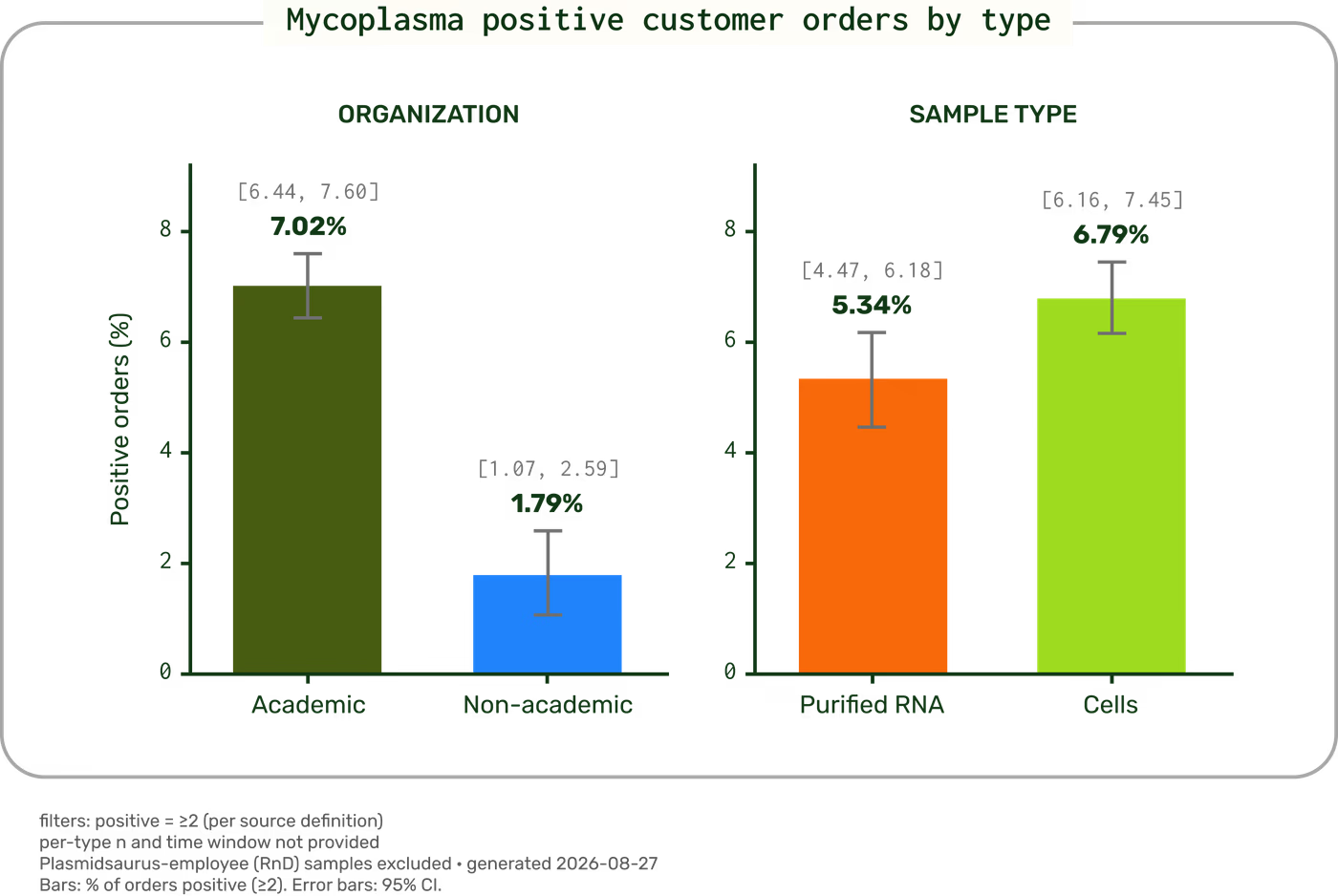
Across the samples we processed, we detected mycoplasma in more than 7% of academic orders and ~2% of non-academic orders, and more than 5% of purified RNA and more than 6% of cell submissions.
We measured the accuracy of our screen using a commercially available qPCR kit. Briefly, we identified a subset of 280 samples that our screen detected as mycoplasma positive, in addition to 401 matched screen-negative samples. We then purified gDNA and performed the qPCR according to the manufacturer's instructions.
279 of the 280 samples detected as mycoplasma positive in our screen were also positive by qPCR. Among the 401 samples that were mycoplasma negative by our screen, 287 were also negative by qPCR while 114 were positive by qPCR. Comparing our screen to the qPCR results across all samples, this works out to a sensitivity of 71%, a specificity of 99.7%, a positive predictive value of 99.6% and negative predictive value of 71.6%.
| Mycoplasma positive by qPCR | Mycoplasma negative by qPCR | |
|---|---|---|
| Plasmidsaurus screen positive | 279 | 1 |
| Plasmidsaurus screen negative | 114 | 287 |
Overall, our screen is highly specific for mycoplasma, meaning a positive call is highly likely to be true contamination, while a non-call in our screen doesn’t rule out mycoplasma.
Interpreting your results
Our mycoplasma detection screen is designed to detect when mycoplasma is present in a sample, but not to determine when mycoplasma is absent.
Mycoplasma detection reporting requires at least two submitted samples. To minimize false positives, contamination is reported only when two or more samples test positive in a single order.
A positive result means the mycoplasma signal is well above the background we see in uninfected samples, in a range clearly separated from normal. This is a strong indication that mycoplasma is present in your sample, and the gene expression data should be regenerated.
A negative result is less conclusive. The absence of detection does not rule out an infection. A sample that falls below our reporting threshold has not produced enough high-confidence reads to call a detection. This can happen for several reasons: a very low infection load, a species outside our reference set, or sample-level factors (low input, degraded RNA, etc.) that reduce the number of non-host reads available for screening.
For these reasons, our mycoplasma detection screen is not a replacement for routine mycoplasma testing.
Mycoplasma screen methods
After we map your RNA-Seq reads to the host reference that you selected as part of your order (e.g. human, mouse), some reads will not align. We take these unmapped reads and align them against a curated selection of mycoplasma genomes covering nine species: the eight most commonly reported as cell culture contaminants, plus one additional species we identified in customer data. After stringent filtering for alignment identity and overall quality, we count the number of reads that cleanly and confidently align to each species.
Once we know the number of reads that align to any of these nine species, we use a dynamic threshold to determine whether mycoplasma reads were detected. The threshold sits above the noise floor we observe in clean samples while remaining sensitive to genuine infections.
What we can detect
Our analysis currently screens for nine species: the eight mycoplasma and related species most frequently reported in cell-culture contaminants, plus one additional species we identified in customer data.
Most commonly reported in the literature:
- Acholeplasma laidlawii
- Mesomycoplasma hyorhinis
- Metamycoplasma salivarium
- Mycoplasma orale
- Mycoplasma hominis
- Mycoplasma pirum
- Mycoplasmopsis fermentans
- Mycoplasmopsis arginini
Identified in customer data:
- Mycoplasma yeatsii
Recommendations following a positive result
We recommend taking several actions if your RNA-Seq samples are found to be positive for mycoplasma. The safest course of action is to dispose of any cultures that are growing from the same stock that was used to generate the data and to regenerate the data using new, uninfected cell lines. The steps below are best practices. For detailed protocols, consult a cell-culture repository like ATCC.
Initial response:
- Discard all infected cell cultures growing from the cell lines used to generate your RNA-Seq data.
- Discard any media and serum used to propagate the infected lines.
- Decontaminate the workspace. Disinfect surfaces, biosafety cabinets, incubators (including water pans), and equipment.
- Thaw and test your cryopreserved stock using an orthogonal method to determine whether it was contaminated. If it is mycoplasma negative, use it to repeat your experiments.
- If your frozen stocks are also contaminated, discard them. Obtain uninfected cells from a repository such as ATCC. If the line isn't commercially available, identify another mycoplasma-free source.
- If the line is irreplaceable, you may consider elimination with a mycoplasma removal agent (MRA) such as Plasmocin. MRAs are broad-spectrum antibiotics added to the culture; treatment can take weeks to months depending on the severity of the infection, and cannot guarantee complete removal.
- Institute routine mycoplasma testing of all cell lines in your lab, and test with a fast screen such as PCR before performing any gene expression analysis.
Think carefully about the existing RNA-Seq data, including historical data that used the same lines or that was generated at the same time these lines were cultured in your lab. An infection will have affected your results. Data may still be informative but should be reported transparently.
Summary
Mycoplasma is a common, easily missed contaminant that can invisibly distort RNA-Seq results, which is why we screen samples automatically as part of our standard pipeline. Our screen aligns non-host reads against nine species: the eight reported to be responsible for most cell-culture contamination plus one more we identified in customer data. The pipeline flags a detection when the signal rises well above the background of uninfected samples. The result is asymmetric: a positive result is a strong indication that your sample is contaminated and the gene expression data has been impacted, while a negative result does not rule out a low-level infection. For this reason, our mycoplasma results complement but do not replace routine mycoplasma testing.
References
ATCC. Mycoplasma contamination. ATCC (accessed 27 August 2026). https://www.atcc.org/the-science/authentication/mycoplasma-contamination
Borchsenius, S.N., Vishnyakov, I.E., Chernova, O.A., Chernov, V.M. & Barlev, N.A. Effects of mycoplasmas on the host cell signaling pathways. Pathogens. 9: 308 (2020). doi: 10.3390/pathogens9040308
Olarerin-George, A.O. & Hogenesch, J.B. Assessing the prevalence of mycoplasma contamination in cell culture via a survey of NCBI's RNA-seq archive. Nucleic Acids Res. 43: 2535–2542 (2015). doi: 10.1093/nar/gkv136
PS-0004-E | v1.2 | Revised Sept. 10, 2026